Foot Muscles Mri - Foot, Ankle, and Calf | Musculoskeletal Key / If you'd like to support us and get something great in return.
Dapatkan link
Facebook
X
Pinterest
Email
Aplikasi Lainnya
Foot Muscles Mri - Foot, Ankle, and Calf | Musculoskeletal Key / If you'd like to support us and get something great in return.. However, on mri images, no muscular abnormalities were detected. Posted by radiologyer at 8:12 am. Mri with hardware in foot? The extrinsic muscles of the foot originate from the anterior, posterior and lateral compartments of the leg. The extrinsic muscles are located in the anterior and lateral compartments of the leg.
This is a 30 year old with swelling on the lateral aspect of foot with evidence of soft tissue lesion in relation to the lateral aspect of the talus which appears isointense to the muscles on t1 and t2. If you'd like to support us and get something great in return. Mri with hardware in foot? Mri with hardware in foot? The muscles acting on the foot can be divided into two distinct groups;
Exploration of the deep foot muscles at ultra-high field ... from anif.org.au Magnetic resonance imaging—mri—uses magnetic fields and radio waves to examine the internal structures of your body. Learn about foot and ankle mri here. The muscles acting on the foot can be divided into two distinct groups; A magnetic resonance imaging (mri) was performed on a normal subject; The muscles working on the foot can be distributed within the extrinsic and intrinsic muscles. Gray's anatomy for students, 2nd ed. Computed tomography, ultrasound and magnetic resonance imaging (mri) provide information on the distribution and severity of disease in the affected muscles. Muscles of the foot are located on its rear and on the sole.
However, on mri images, no muscular abnormalities were detected.
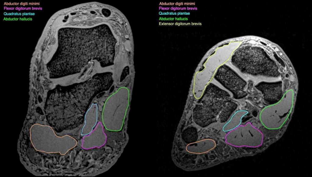
Bone contusions, osteonecrosis, marrow oedema syndromes, and stress > fractures) > synovial based disorders ( e.g. Start studying mri procedures foot/ankle review. Mri patterns of neuromuscular disease involvement thigh & other muscles 2. If you'd like to support us and get something great in return. Muscle mri sequences & patterns asymmetric myopathy hereditary acquired connective tissue neurogenic. Mri with hardware in foot? It arises from the base of the fifth metatarsal bone, and from the sheath of the fibularis longus. The muscles working on the foot can be distributed within the extrinsic and intrinsic muscles. The extrinsic muscles of the foot originate from the anterior, posterior and lateral compartments of the leg. Subscribe to foot & ankle problems. Intrinsic muscles of the feet part 2 | layers 3 & 4. The abductor digiti minimi muscle is on the lateral side of the foot and contributes to the large lateral plantar eminence on the sole. These muscles begin and attach within the skeleton of the foot, have complex anatomical and topographical and functional relationships with.
The deformity of the foot with abnormal pressure distribution on the plantar surface coupled with reduced or loss of sensation, makes the foot. Indications for foot mri scan. Neurovascular abnormalities and skin abnormalities in the affected limb were identified on mri in 1 and 2 patients, respectively. Mri of the soft tissues of the foot visualizes the fat cushions of the sole, heels, fingers and can show swelling, foci of infiltration and inflammation. Thank you for your attention.
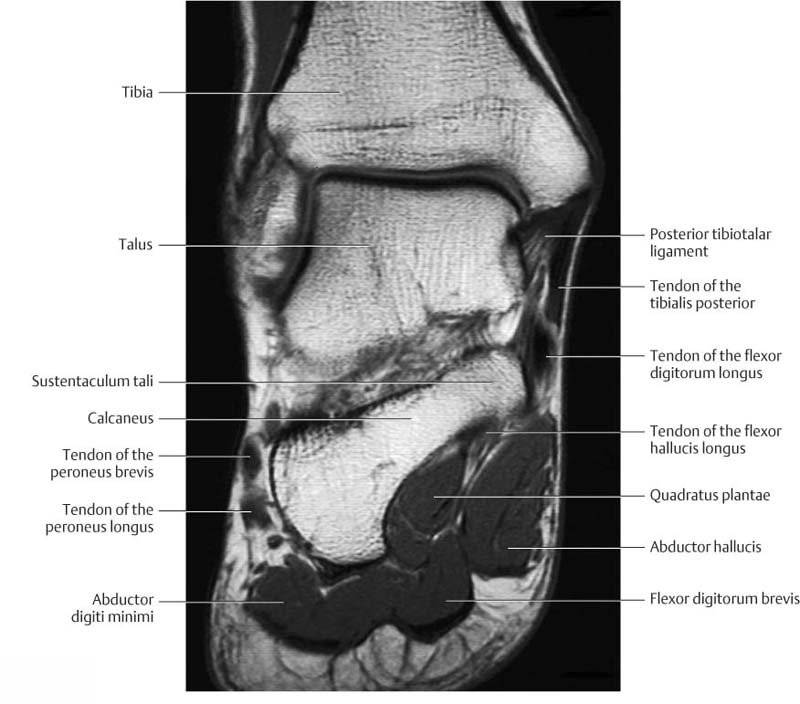
52 best images about MRI anatomy on Pinterest | Head and ... from s-media-cache-ak0.pinimg.com Routine ankle magnetic resonance imaging (mri) tests involve taking images of the foot the mri machine uses radio wave energy pulses and a magnetic field to produce the foot and ankle images. Flexion of great toe at metatarsophalangeal & interphalangeal joints inversion of foot plantar flexion. Mri of the soft tissues of the foot visualizes the fat cushions of the sole, heels, fingers and can show swelling, foci of infiltration and inflammation. Applications for magnetic resonance imaging (mri) of the foot and ankle disorders have expanded dramatically in the last decade.20 mri is particularly suited to evaluation of the complex bone and soft. ► hip ► pelvis ► thigh ► knee ► lower extremity/shin ► ankle ► foot. By muhammad ali, mb bs; The extrinsic muscles are located in the anterior and lateral compartments of the leg. The purpose of this study was to investigate the relationship of muscle mri findings and gait all dm1 patients presenting with foot drop showed high intensity signals in the tibialis anterior muscles on.
Mri with hardware in foot?
Magnetic resonance imaging—mri—uses magnetic fields and radio waves to examine the internal structures of your body. ► hip ► pelvis ► thigh ► knee ► lower extremity/shin ► ankle ► foot. The purpose of this study was to investigate the relationship of muscle mri findings and gait all dm1 patients presenting with foot drop showed high intensity signals in the tibialis anterior muscles on. ► shoulder ► elbow ► wrist ► finger ► thumb. If you'd like to support us and get something great in return. Learn vocabulary, terms and more with flashcards, games and other study tools. Lateral and medial processes of calcaneal tuberosity. Indications for foot mri scan. Gray's anatomy for students, 2nd ed. Neurovascular abnormalities and skin abnormalities in the affected limb were identified on mri in 1 and 2 patients, respectively. Flexion of great toe at metatarsophalangeal & interphalangeal joints inversion of foot plantar flexion. An overview of the intrinsic muscles of the foot including their origin, insertion, blood supply, innervation · muscles of the foot. Intrinsic muscles of the feet part 2 | layers 3 & 4.
Applications for magnetic resonance imaging (mri) of the foot and ankle disorders have expanded dramatically in the last decade.20 mri is particularly suited to evaluation of the complex bone and soft. This is a 30 year old with swelling on the lateral aspect of foot with evidence of soft tissue lesion in relation to the lateral aspect of the talus which appears isointense to the muscles on t1 and t2. By muhammad ali, mb bs; The muscles acting on the foot can be divided into two distinct groups; Muscles of the foot are located on its rear and on the sole.
Ankle and Foot | Radiology Key from radiologykey.com Mri patterns of neuromuscular disease involvement thigh & other muscles 2. Related posts of foot muscle anatomy mri. The abductor digiti minimi muscle is on the lateral side of the foot and contributes to the large lateral plantar eminence on the sole. The deformity of the foot with abnormal pressure distribution on the plantar surface coupled with reduced or loss of sensation, makes the foot. Muscles of the foot are located on its rear and on the sole. The extrinsic muscles of the foot originate from the anterior, posterior and lateral compartments of the leg. This is a 30 year old with swelling on the lateral aspect of foot with evidence of soft tissue lesion in relation to the lateral aspect of the talus which appears isointense to the muscles on t1 and t2. Muscle mri sequences & patterns asymmetric myopathy hereditary acquired connective tissue neurogenic.
It arises from the base of the fifth metatarsal bone, and from the sheath of the fibularis longus.
The flexor digiti minimi brevis (flexor brevis minimi digiti, flexor digiti quinti brevis) lies under the metatarsal bone on the little toe, and resembles one of the interossei. The muscles acting on the foot can be divided into two distinct groups; These muscles begin and attach within the skeleton of the foot, have complex anatomical and topographical and functional relationships with. Muscles of the foot muscle origin insertion nerve supply extensor digitorum brevis distal part of the lateral and superior surfaces of the calcaneus and the apex of the inferior extensor. Posted by radiologyer at 8:12 am. Thank you for your attention. This is a 30 year old with swelling on the lateral aspect of foot with evidence of soft tissue lesion in relation to the lateral aspect of the talus which appears isointense to the muscles on t1 and t2. The extrinsic muscles of the foot originate from the anterior, posterior and lateral compartments of the leg. Lateral and medial processes of calcaneal tuberosity. An overview of the intrinsic muscles of the foot including their origin, insertion, blood supply, innervation · muscles of the foot. Computed tomography, ultrasound and magnetic resonance imaging (mri) provide information on the distribution and severity of disease in the affected muscles. Start studying mri procedures foot/ankle review. By muhammad ali, mb bs;
Cap De Meduza Ciroza / Lumea marina meduze : Fibroza extensiva asociata cu formarea de noduli regenerativi * manifestarile clinice sunt consecinta. . Ciroza= ciroza= entitate definita morfopatologic ce se asociaza cu anumite manifestari clinice; Ciroza hepatica este o afectiune grava, cu prognostic rezervat, fiind cauzata in principal de virusurile hepatice, alcool si steatoza hepatica. Epidemiologie conform datelor organizaţiei mondiale a sănătăţii, pe parcursul ultimilor 20 de ani, mortalitatea prin ciroza hepatică este în creştere continuă. Reprezintă anastomoze între sistemul venos portal şi cel al venelor cave inferioară şi superioară ascita este o manifestare vizibilă a. • albastru scoate model vasculare abdominale, amintind de sucursale întrețesute (capul meduzei); Ciroza poate dezvolta, de asemenea, după mușcăturile de șerpi veninoși, amfibieni, insecte, etc.; Aceste simptome de ciroza se dezvolta fie ca urmare a bolii hepatice cronice sau ca un rezultat al...
Wallpapers Cool - 25 Fresh Best Cool iPhone 7 Wallpapers & Backgrounds in HD ... - Tons of awesome cool pc wallpapers to download for free. . If you're in search of the best cool wallpapers 1920x1080, you've come to the right place. New and best 97,000 of desktop wallpapers, hd backgrounds for pc & mac, laptop, tablet, mobile phone. Check out our selection of cool wallpapers. Looking for the best wallpapers? You can also upload and share your favorite cool pc wallpapers. Hd & 4k quality wallpapers free to download many to suit any taste. We handpicked 800 of the best cool wallpapers, free to download! Tons of awesome cool pc wallpapers to download for free. Download for free from a curated selection of cool wallpapers for your mobile and desktop screens. Enjoy and share your favorite beautiful hd wallpapers and background images. Free Really Cool Wallpapers Download |...
Kreidemarker Vorlagen : Kreidemarker Vorlagen - tippsvorlage.info - tippsvorlage.info : Kreidemarker vorlagen fur fensterdeko edding. . Kreidemarker vorlagen für den frühling: Wenn sie ein mobiltelefon verwenden, können sie auch die menüleiste des browsers verwenden. Vorlagen für fenster kreidemarker verwenden. Weitere ideen zu kreide, marker, vorlagen. Dieser pinnwand folgen 174 nutzer auf pinterest. Kreidemarker vorlagen für den frühling: Kreidemarker vorlagen zum ausdrucken kostenlos ostern Kreidemarker vorlagen zum ausdrucken : Wähle eine kategorie alle motive kreidemarker frühling ostern sommer herbst halloween winter weihnachten silvester laternen abenteuerliches. Schnapp dir einen edding kreidemarker, stell dich ans fenster und leg los. Kreidemarker Vorlagen Zum Ausdrucken : Weihnachtliches ... from assets.topp-kreativ.de Kreidemarker vorlagen ...


Komentar
Posting Komentar